The effect of using kefir grains and mesenchymal stem cells in LPS-induced Alzheimer’s disease neuroinflammatory model
- Inicio
- Comité Editorial
- Lineamientos
- Carta de Cesión de Derechos
- Información Legal
- Acerca de la Revista
- Bases de Datos
- Contacto
- ISSN 2007-3054
- Centro de Investigaciones Cerebrales
Universidad Veracruzana
Mai M. Anwar1*, Ola S. M. Ali2, Laila Ahmed R.3, Badawi A. M.1, Nadia A. Eltablawy1
1Department of Biochemistry, National Organization for Drug Control and Research (NODCAR), Egypt. 2Department of Biochemistry, Faculty of Pharmacy, Al-Azhar University, Egypt. 3Department of Biochemistry and Molecular Biology, Faculty of Medicine, Cairo University, Egypt
Resumen/Abstract
Introduction
Materials and Methods
Biochemical analysis
Statistical analysis
Histopathological examination of brain tissues
Discussion
Conclusion
Abbreviations
Ethics approval and consent to participate
Consent for publication
Availability of data and materials
Competing interests
Funding
Authors’ contributions
Acknowledgements
References
Mail
Background: Alzheimer’s disease (AD) is characterized by severe accumulation of amyloid plaques and neurofibrillary tangles accompanied with cognitive dysfunction leading to major changes affecting the quality of patient daily life pattern. Objective: To investigate the effect of MSCs and/or milk kefir grains in LPS –induced AD in female albino rats in alternating manner. Materials and Methods: Sixty female albino rats were divided into equal six groups (ten rats each): group 1: healthy control; group 2: LPS-induced AD; group 3: LPS-induced AD rats received single intravenous injection of MSCs; group 4: LPS-induced AD rats received oral milk kefir; group 5: LPS-induced AD rats received a single intravenous injection of MSCs with a daily milk kefir grain administration for a month; group 6: Rats received kefir for one week prior to the induction of AD followed by a single intravenous injection of MSCs with a daily milk kefir grain administration for a month. AD was assessed by T maze behavioural test month after induction. Brain tissue was collected for monitoring BDNF, Bax, Bcl-2 and seladin-1 gene expression with the measurement of TNF-α, IL-10 and tissue cholesterol. Plasma lipid profile, GSH and MDA were also determined. Results: Revealed that significant elevation of lipid profile and oxidative stress in association with LPS-induction. BDNF, Bcl-2 and seladin-1 gene expression were significantly reduced in AD while Bax mRNA gene was significantly increased. Administration of kefir and /or MSCs suppressed LPS –induced AD. Conclusion: The pre and co-administration of kefir with MSCs could act as a potent modulator attenuating the underling pathological inflammatory process accompanying AD which results in the progression of brain damage.
Keywords: Alzheimer's disease, MSCs, kefir grains, LPS, Bax, BDNF, Seladin-1.
La enfermedad de Alzheimer (EA) se caracteriza por una acumulación severa de placas amiloides y ovillos neurofibrilares acompañados de disfunciones cognitivas severas que conducen a cambios importantes que afectan la calidad del patrón de vida diaria del paciente. El objetivo del presente estudio es investigar los efectos de la administración de células madre mesenquimales (MSC) y/o granos de kéfir de leche en el modelo de tipo neuroinflamatorio de EA inducida por LPS de manera alternativa. Se observó que una elevación significativa del perfil lipídico y el estrés oxidativo estaban relacionados con EA de tipo neuroinflamatoria inducida con LPS. La expresión del gen BDNF, Bcl-2 y seladin-1 también se redujo significativamente en ratas con EA, mientras que la expresión relativa de Bax aumentó significativamente. La administración de granos de kéfir de leche y/o MSC suprimió los inconvenientes de la EA incluyendo cambios de comportamiento y memoria. Conclusión: La administración previa y la coadministración de granos de kéfir de leche con MSC pueden actuar como un neuromodulador activo que atenúa el proceso inflamatorio patológico subyacente que acompaña a la EA, lo que resulta en la progresión de daños en el tejido cerebral.
Palabras clave: Enfermedad de Alzheimer, neuroinflamación, MSC, granos de kéfir, LPS, Bax, BDNF, Seladin-1.
Alzheimer’s disease (AD) is a progressive neurodegenerative disorder and the most common form of dementia in the world.1 Clinically; AD is typically characterized by progressive memory loss, impairment of other cognitive functions and inability to perform activities of daily living. The earliest clinical stage of AD is defined as mild cognitive impairment (MCI) and it is characterized by progressive memory impairment and /or others cognitive functional abilities. Nowadays, the available chemical therapeutic agents are only able to slow down the AD progressive drawbacks.2 Pathologically; AD is characterized by two highlighted protein abnormalities: extracellular amyloid-β (Aβ) deposition and intra-cellular neurofibrillary tangles (NFT) formation, leading to extensive neuronal damage. Aβ peptides derive from cleavage of membrane amyloid precursor protein (APP) by beta- APP cleaving enzyme 1 (BACE1) and the γ-secretase complex containing the complex presenilin protein (PSEN) while The NFT is usually derived from abnormal aggregation of hyperphosphorylated microtubule associated with tau protein.3-7
Inflammation has been likely associated with Alzheimer’s disease pathogenesis for over a decade with increasing evidence suggesting that abnormalities in cholesterol homeostasis could have an important role as well.8 Central Nervous System (CNS) inflammation in AD is characterized by reactive microglia M1-type, elevated IL-1 and other complement factors.9 Aβ aggregates can stimulate oxidation in several ways including localized H2O2 production10 and free radical11 Both oxidative damage,12 and inflammation9,10 can be considered as a pre-disposing factor for plaques accumulation and neurodegeneration disorders. The therapeutic potentials of stem cells in several brain disorders are driving researchers to apply stem cell-based therapies as a progressive way of combating neurodegenerative diseases and neuroinflammatoin as well.12-14 Neural stem cells have been proved to rescue memory impa-irment in AD models by releasing brain-derived neurotropic factor (BDNF).15 Also, bone marrow derived mesenchymal-mal Stem Cells (BM-MSCs) modulate anti-inflammatory im-mune response with alleviated Aβ aggregations and memory deficits in AD animals model.16
As we mentioned, neurodegenerative diseases including AD are devastating disease that can be initiated from different stated conditions including inflammatory triggering factors.17 Lipopolysaccharides (LPS) is the outer membrane of gram-negative bacteria which can be considered as one of the main stated AD neuroinflammatory initiatory factors in different subjects including rats by targeting toll-like receptor (TLR-4) leading to the propagation of series of inflammatory cascades using different downstream adaptors molecules and ending with a sever neuroinflammatoin in brain tissue resulting in the formation of AD neuro-inflammatory type with enhanced Amyloid β and tangles formation.18,19 These inflame-matory changes will result in behavior modifications on LPS subjected models including sickness behavior and dizziness as a result of exaggerated cytokines production and neurodegenerative cascading process.20,21
kefir is a kind of natural beverage consisting of gelatinous and irregular grains formed by a consortium of yeasts and lactic acid bacteria causing acid-alcoholic fermentation in sugar and milk solutions with a consistency thicker than milk and lighter than yoghurt.22 These micro flora are embedded in a resilient polysaccharide matrix, called Kefiran, composed of branched chains of glucose and galactose as metabolic products of souring milk lactose and other byproducts.23 There are a number of approximately more than 50 bacteria and yeast strains living together in kefir grains. This number may vary depending on the culture media used and the country of the grains source.24 kefir can also be considered as a probiotic continual source due it’s varies beneficial effects besides its nutritional actions. There are several studies investigating the immune-modulatory-linked actions, anti-inflammatory effects, pathogenic barrier actions, anti-diabetic and digestive effects abilities by a regular daily kefir intake.22
The aim of this study was to investigate the effects of MSCs and/or kefir in Alzheimer’s disease induced in female albino rats and to demonstrate their efficiency in combating AD neuroinflammatory drawbacks whether administered in combination or even alone. Studying the action of probiotics represented in the form of milk kefir grains on relieving the drawbacks of Alzheimer’s disease and their ability to improve memory related dysfunction was also the core of establishing this study.
2.1. Preparation of bone marrow (BM) derived mesenchymal stem cells from rats
Bone marrow was harvested by flushing the tibiae and femurs of 8 weeks female white albino rats with Dulbecco’s modified Eagle’s medium (DMEM, GIBCO/BRL) supplemented with 10% fetal bovine serum (FBS) (GIBCO/BRL). Nucleated cells were isolated with a density gradient [Ficoll/Paque (Pharmacia)] and re-suspended in complete culture medium supplemented with 1% penicillin–streptomycin (GIBCO/BRL). Cells were incubated at 37°C in 5% humidified CO2 for 15 days as primary culture or upon formation of large colonies. When large colonies developed, cultures were washed twice with phosphate buffer saline (pH 7) and the cells were trypsinized with 0.25% trypsin in 1mM EDTA (GIBCO/BRL) for 5 min at 37°C. After centrifugation, cells were re-suspended in serum supplemented medium and incubated in 50 cm2 culture flasks (Falcon). The resulting cultures were referred to as first-passage cultures (Alhadlaq and Jeremy, 2004). Cells were identified as being MSCs by their morphology, adherence and their differentiation power to osteocytes and chondrocytes.25
2.2. Preparation of kefir
Milk kefir grains were used as a probiotic live active culture grains in organic powdered skimmed milk purchased from Cultures for Health, USA (17978S, Grasle Rd. Ore City OR 97045). Freeze dried kefir grains (20mg) were inoculated to 100 ml of pasteurized milk. All grains inoculated milk was incubated at 20°C for 24 h. At the end of fermentation, the milk was filtered to remove kefir grains with a daily repeating of this step till the end of experiment. The milk kefir grains were then freeze-dried and stored at 4°C until required.26
2.3. Animals and experimental design
The study was carried out using sixty female albino rats weighing 200-250g. Rats were bred and maintained in air-conditioned animal house with specific pathogen-free conditions and were subjected to a 12:12-h day light/darkness with unlimited access to food and water. All the ethical protocols for animal treatment were followed and supervised by the animal facilities, Faculty of Medicine, Cairo University. All the animals included in the experiment received approval from the Institutional Animal Ethics Committee. To achieve the ultimate goal of this study, animals were randomized divided into equal six groups (Ten rats each):
Group I (control): Control group.
Group II (AD positive control): Rats of this group were injected intraperitoneally (I.P) with 0.56mg/kg body weight of LPS dissolved in 1 ml of sterile PBS (PH7) to induce neuro-inflammatory AD type as a modified single dose previously published.27
Group III (AD+MSCs): Lipopolysaccharides (LPS) induced neuroinflammatory AD type in rats which received MSCs (which were processed and cultured for 14 days, in a single dose of 1μL (500 x103/μL) cells.28
Group IV (AD + kefir): LPS-induced neuroinflammatory AD type in rats received 3.5ml/kg body weight of milk kefir grains by oral gavage once daily for a month as a modified previous dose response previously published.26
Group V (AD+MSCs+kefir): LPS-induced neuroinflammatory AD type in rats received a single intravenous injection (IV) of MSCs simultaneously with milk kefir grains once daily for a month as mentioned above.
Group VI (kefir+AD+MSCs+kefir): Rats of this group received milk kefir grains for one week prior to the induction of neuroinflammatory AD type by LPS followed by single intravenous Infusion of MSCs simultaneously with daily milk kefir administration for a month.
2.4. Behavior study through time consumed in T-maze test
Behavior study was done one day after the last dose of the given treatments. Rats were transported to the testing facility (Behavioral Lab, Faculty of Medicine, Cairo University), 30 minutes period was allowed prior to testing to adapt to the environment and to minimize the effect of stress due to transfer.
2.5. Apparatus setting and animal preparation
The automatic modified T-maze apparatus was constructed of white plastic runways with 25cm high walls and was divided into 6 areas (A1, A2, S1, S2, P1, P2) by sliding doors (s1, s2, s3, a1, a2, p1, p2. The stem of the T is composed of two arms. The end of each arm is equipped with a pellet sucrose dispenser (20 mg, Formula 5 TUT, Test Diet, Richmond, IN, USA) as a reward. Two rats per cage in a temperature-controlled room (23±2°C) with a 12h light/dark cycle, according to guidance and protocols established by local Animal Care and Use Committee. Transfer all the cages containing rats into the soundproof room from the housing room at least thirty min before the first trial begins. All the experiments should be always performed during the same time period (e.g., 9:00 AM to 6:00 PM) with slight modification of.29 Pre-training sessions are required followed by either a forced alternation task or left-right discrimination task.
2.6. Forced alternation task
In the forced alternation task, each trial consists of a forced choice run followed by a free choice run. The rat is allowed to enter the area and to consume the pellet. When the rat has eaten the pellet, the door near the food tray of the arm that the rat currently stays is opened. Then, the rat approaches the door neighboring the start box, so it can return to the start box. Following the forced-choice run, the free-choice run begins. The rat is allowed to choose between the two arms. If the rat enters the opposite arm that it was forced to choose in the forced-choice run, its response is considered to be "Correct" and the rat receives a sucrose pellet. If the rat goes to the same arm as that visited in the forced-choice task, the rat is opened, and the rat can return to the start box. In these trials, time consumed for each rat in each session was measured with slight modification.29
At the end of the experiment, animals were anesthetized with sodium phenobarbital (60 mg/kg), blood samples were collected in heparinized tubes from the retro-orbital venous plexus and plasma were carried out by using cooling centrifugation at 3000 rpm for 10 min and stored at -20 °C until use. After collection of blood samples, all animals were sacrificed by decapitation and brains were rapidly removed and cut into 2 symmetrical halves by midline incision. One part was removed and immediately immersed in 10% buffered formalin for histopathological examinations and the other half was stored at –80°C for further biochemical analysis.
3.1. Preparation of brain tissue homogenate
The brain tissue was homogenized in 10 % PBS buffer (pH 7) for 10 min and centrifuged for 20 min at 4,000 rpm. The clear supernatant obtained was then divided into three portions and were kept frozen at -80 °C till analysis.
3.2. Real-time quantitative analysis for BDNF, Bax, Bcl-2 and seladin1 gene expression
Brain total RNA was extracted from the homogenate using RNeasy purification reagent (Qiagen, Valencia, CA), cDNA was generated from 5 μg of total RNA extracted with 1 μl (20 pmol) antisense primer and 0.8 μl superscript AMV reverse transcriptase for 60 min at 37 °C. The relative abundance of mRNA species was assessed on an ABI prism 7500 sequence detector system (Applied Biosys-tems, Foster City, CA). PCR primers were designed with Gene Runner Software (Hasting Software, Inc., Hasting, NY) from RNA sequences from Gene Bank. Primers were checked for specificity by melting curve analysis (MCA). Melting curve analysis (MCA) is a kind assessment for ensuring the double-stranded DNA dissociation produced from the qPCR reaction during heating, and also used in qPCR reactions with intercalating dyes including SYBR Green which only fluoresces when it is in double-stranded bonded DNA form. All primer sets had a calculated annealing temperature of 60°. Quantitative real time-PCR was performed in duplicate in a 25-μl reaction volume consisting of 2X SYBR Green PCR Master Mix (Applied Biosystems), 900 nM of each primer and 2–3 μl of cDNA. Amplification conditions were 2 min at 50°, 10 min at 95° and 40 cycles of denaturation for 15 s and annealing/extension at 60° for 10 min. Data from real- time assays were calculated using the v1•7 sequence detection software from PE. Bio- systems (Foster City, CA). Relative expression was calculated using the comparative Ct method as previously described. All values were normalized to the B-actin gene and reported as fold change over background levels detected in AD.
3.3. Determination of IL-10 and TNF-α level in brain tissue
The second portion of Brain Homogenate were used in the determination of tissue interleukin -10 (IL-10) and tumor necrosis factor-alpha (TNF-α) by commercially available Enzyme-linked immunosorbent assays (ELISA) kits supplied from Q&D system Quatin USA according to the manufacturer`s instructions. Protein concentrations were determined with bovine serum albumin as a standard.30
3.4. Determination of brain tissue cholesterol level
The third portion of Brain Homogenate was used in the determination of tissue total cholesterol level;26 Kit was supplied from Cell Bio Labs. Protein concentrations were deter-mined with bovine serum albumin as a standard.
3.5. Determination of plasma lipid profile
Plasma cholesterol31 , triglycerides32 and HDL-CHO33 were performed using a commercial kits supplied from Cell Bio Labs. Meanwhile, LDL-CHO level was calculated.34
3.6. Determination of blood GSH and MDA
Blood GSH35 and MDA36 were performed using commercial kits that were obtained from Biodiagnostic, Egypt.
3.7. Analysis of brain histopathology
The obtained tissue sections were collected on glass slides, deparaffinized and stained by hematoxylin and eosin (H&E). Examination was performed by light microscopy.37
Data were expressed as mean ± SE. Significant differences were determined by using One Way Anova and post-hoc tests (Duncan’s multiple comparison test). p< 0.05 was considered significant. Data were statistically analyzed using the statistical package for social science software version18 (SPSS, Chicago, IL, USA).
4.1. Behavior study through time consumed in T- maze test
In the current investigation, administration of LPS (0.56 mg/kg) resulted in a significant increase in the time spent by the rats when compared with the negative control group. Treatment with MSCs and/or milk kefir grains resulted in a significant protection against LPS induced changes (p <0.05) and significantly decreased the time spent by rats in T-maze test. The greatest decrease in time spent by rats was seen in the prophylactic group VI (kefir+AD+MSCs+kefir) indicating a major memory recovery. All Groups were signific-antly different from control values (p < 0.05) as shown in Figure1.
4.2. Effects of MSCs and/or milk kefir on BDNF, Bax, Bcl-2 and seladin-1 relative expression on brain tissue
Concerning gene relative expression, there was a major significant decrease in BDNF, Bcl-2, seladine-1 with an increase in Bax relative expression of the LPS-induced AD when compared with the control group (P<0.05) as shown in Table 1. Following MSCs injection and /or milk kefir grains administration, there was an improvement in the reduction level of brain tissue BDNF, Bcl2, seladine-1 with decrease in Bax relative expression with variations in the percent of change among the groups. Also, a synergistic effect was potentially noticed by the pre and co-administration of milk kefir with MSCs in LPS induced rats as shown in Figures 2a, 2b.
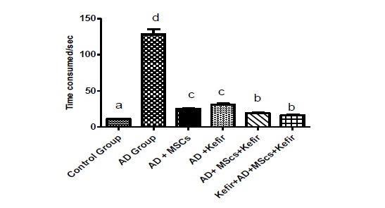
Figure 1: Mean ± SE (n=10) of time consumed in T-maze. Same small letters mean insignificant difference between treated groups using one-way ANOVA and followed by Duncan’s multiple comparison test at p< 0.05.
4.3. Effects of MSCs and /or milk kefir on TNF-α and IL-10 levels of brain tissue
TNF-α and IL-10 level were estimated in brain supernatant of all the six studied groups. As shown in Table 2 and Figures 3a and 3b. Levels of TNF-α were significantly increased with a simultaneous decrease in IL-10 level of the LPS –induced AD when compared to control group (p < 0.05), indicating that LPS induced a neuroinflammatory process which may contribute to AD.
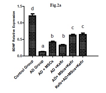
Figure (2a): Mean ± SE (n=10) of MSCs and/or milk kefir grains administration on brain BDNF relative expression of LPS- induced AD neuroinflammatory type in albino rats. Same small letters mean insignificant difference between treated groups using one-way ANOVA and followed by Duncan’s multiple comparison test at p< 0.05
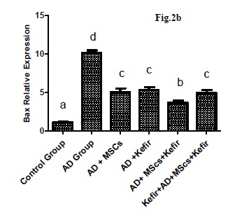
Figure (2b): Mean ± SE (n=10) of MSCs and/or milk kefir grains administration on brain bax relative expression of LPS- induced AD neuroinflammatory type in albino rats. Same small letters mean insignificant difference between treated groups using one-way ANOVA and followed by Duncan’s multiple comparison test at p< 0.05.
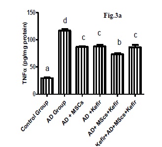
Figure (3a): Mean ± SE (n=10) of MSCs and/or milk kefir grains administration on brain tissue TNF-α (pg/mg protein) of LPS-induced AD neuroinflammatory type in albino rats. Same small letters mean insignificant difference between treated groups using one-way ANOVA and followed by Duncan’s multiple comparison test at p< 0.05.
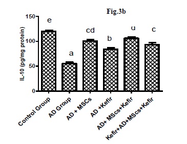
Figure (3b): Mean ± SE (n=10) of MSCs and/or milk kefir grains administration on brain tissue IL-10 (pg/mg protein) of LPS- induced AD neuroinflammatory type in albino rats. Same small letters mean insignificant difference between treated groups using one-way ANOVA and followed by Duncan’s multiple comparison test at p< 0.05.
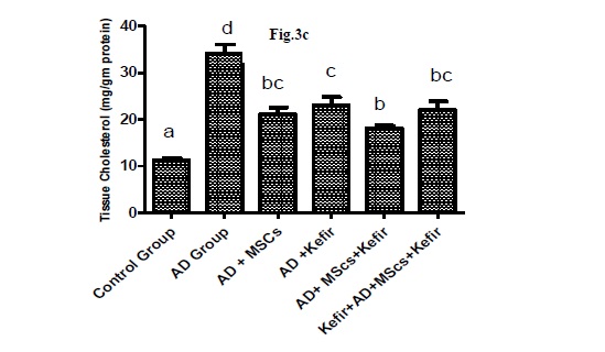
Figure (3c): Mean ± SE (n=10) of MSCs and/or milk kefir grains administration on brain tissue cholesterol level of LPS-induced AD neuroinflammatory type in Albino rats. Same small letters mean insignificant difference between treated groups using one-way ANOVA and followed by Duncan’s multiple comparison test at p< 0.05.
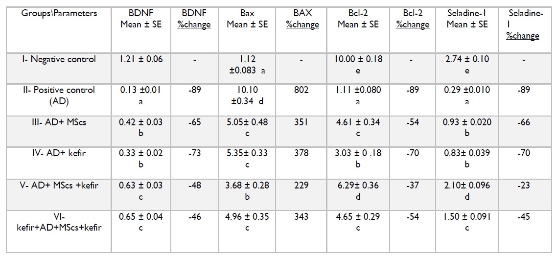
Table 1: Represents the mean of 10 rat’s ± SE. Same small letters mean insignificant difference between treated groups using one-way ANOVA at (p>0.05) followed by Duncan-multiple comparison test. The % of change is corresponding to the negative control Group.
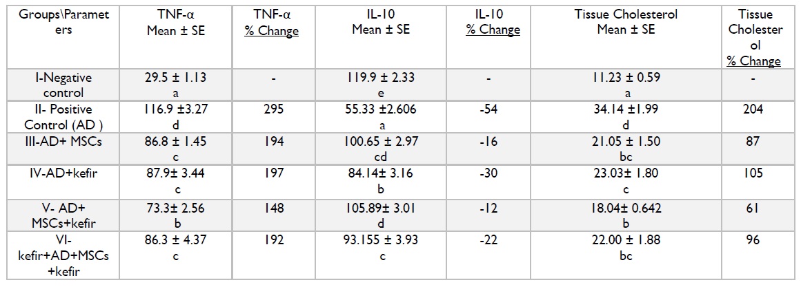
Table 2: Represents the mean of 10 rat’s ± SE. Same small letters mean insignificant difference between treated groups using one-way ANOVA at (p>0.05) followed by Duncan-multiple comparison test. The % of change is corresponding to the negative control group.
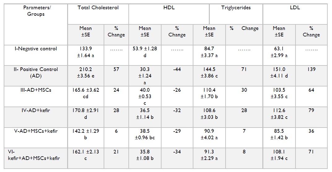
Table 3: Represents the mean of 10 rat’s ± SE. Same small letters mean insignificant difference between treated groups using one-way ANOVA at (p>0.05) followed by Duncan-multiple comparison test. The % of change is corresponding to the negative control Group.to the negative control group.
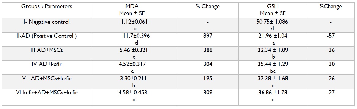
Table 4: Represents the mean of 10 rat’s ± SE. Same small letters mean insignificant difference between treated groups using one-way ANOVA at (p>0.05) followed by Duncan-multiple comparison test. The % of change is corresponding negative control group.
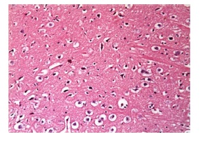
Figure 4: Histopathological examination of control group with normal neuron cells and nerve fibres.
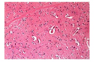
Figure 5: Histopathological examination of LPS-induced neuroinflammatory AD type model showed multiple plaques formed of lamellate fibrils surrounded by multiple apoptotic nuclei.
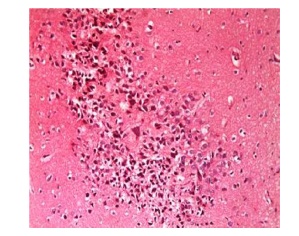
Figure 6: Histopathological examination of LPS-induced neuroinflammatory AD type model with blood vessels plaque formation, apoptotic nuclei and several deposits of (amyloid) material in their wall.
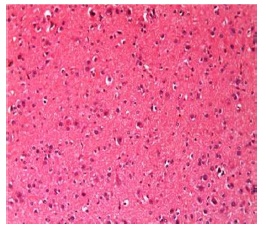
Figure 7: Histopathological examination of LPS-induced neuroinflammatory AD type model treated with MSCs showed less dense plaque formation, apoptotic nuclei and severity of blood vessels congestion.
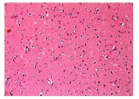
Figure 8: Histopathological examination of LPS-induced neuroinflammatory AD type model treated with milk kefir grains showed slight multiple plaques formation, apoptotic nuclei and slight congestion in blood vessels
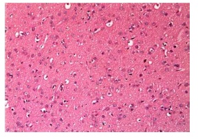
Figure 9: Histopathological examination of LPS-induced neuroinflammatory AD type model treated with both MSCs+milk kefir grains showed slight focal plaques formation, multiple apoptotic nuclei with congestion in blood vessels.
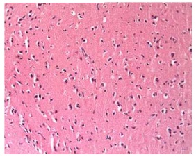
Figure 10: Histopathological examination of prophylactic LPS-induced neuroinflammatory AD type group previously administrated milk kefir grains prior to LPS induction initiation followed by LPS induction then followed by the administration of MSCs+milk kefir grains showed a slight focal plaques formation, no multiple apoptotic nuclei with slight congestion in blood vessels.
4.4. Effects of MSCs and/or milk kefir on cholesterol levels of Brain tissue
The results of the present study show a significant increase in the tissue cholesterol level in the LPS-induced AD group when compared with the negative control with a significant decrease in tissue cholesterol level after the administration of milk kefir grains and/or MScs in different relatives as shown in Table 2 and Figure 3c.
4.5. Effects of MSCs and/or milk kefir on plasma lipid profile
It was shown a significant increase in plasma total cholesterol level, LDL and triacylglycerol with a decrease in the plasma HDL of the LPS-induced AD. kefir and/or MSCs administration cause a decrease in total plasma cholesterol, LDL and triacylglycerol level with an improvement in the reduction level of HDL with variations among the treated groups which indicate the efficacy of milk kefir grains and/or MScs as antihyperlipidemia and anti-hypercholesterolemia.
4.6. Effect of kefir and/or MSCs on plasma lipid peroxidation and reduced glutathione
An observed increase in plasma MDA level associated with a marked decrease in plasma GSH level was showed in LPS-induced AD. Treatment of LPS challenged rats with kefir and /or MSCs produced a significant attenuation in the induced oxidative stress. The obtained results also revealed that the administration of MSCs attenuated the oxidative stress induced by LPS challenge and this attenuating effect was potentially enhanced by the pre and co-administration of milk kefir with MSCs by decreasing the MDA level with improvement in the reduction level of GSH in LPS-induced AD as shown Table 4.
5. Histopathological examination of brain tissues
Concerning the histopathological findings, sections of normal controlled groups showed that the cerebral cortex exhibits normal neurons surrounded by nerve fibers and blood vessels (Figure 4). In LPS induced AD neuroinflammatory rats, an observed multiple plaques formed of lamellated fibrils. Such plaques were surrounded by multiple apoptotic nuclei (Figure 5). Severe inflammation with congested blood vessels was also observed with deposits of amyloid material in their wall and surrounded by extravasated red blood corpuscles when compared with the normal control group (Figure 6). Administration of MSCs showed less dense plaques, multiple and less prominent apoptotic nuclei (Figure 7). On the other hand, administration of milk kefir grains revealed disappearance of multiple plaques, some dense plaques with few apoptotic nuclei (Figure 8). MSCs and milk kefir grains administration lead to slight focal plaques formation, multiple apoptotic nuclei with slight congestion in blood vessels (Figure 9). On the prophylaxis group, there was a slight focal plaques formation, few multiple apoptotic nuclei with less diffused congestion in blood vessels (Figure 10).
Mesenchymal stem cells do have distinguished therapeutic effects via regenerative actions and neuromodulator potentials in addition to their ability to substitute damaged cells and tissues. Bone marrow–derived stem cells types contribute in a unique way in cell turnover with the ability to repair various tissue types, including the brain.38 MSCs are commonly defined as bone marrow derived fibroblast like cells type, which despite the lack of specific surface markers, can be selected by their adherence characteristics invitro and their abilities to differentiate along to three principal mesenchymal lineages osteoblastic, adipocytic and chondrocytic. 39,40 The therapeutic strategy of MSCs includes two directions which to induce the activation of endogenous stem cell and other to regenerate injured damaged tissues through stem cell transplantation. Endogenous stem cells can be induced and can show neuro-protective effects by chemical compounds and factors stimulating secretions such as allopregnanolone (Apα). MSCs also increase neprilysine expression and Aβ-degrading enzyme release leading to the reduction of Aβ deposition, improving memory dysfunction and significant alleviation of AD pathology as a drawback of inhabiting pro inflammatory factors release.41
Kefir is a traditional beverage obtained by the fermentation of kefir grains inoculated in milk containing a wide diversity of lactic and acetic-acid bacteria plus yeasts. 42-43 This kind of unique grains are characterized by inheriting a highly nutritional value as a source of proteins and calcium with being considered as a potent ant-inflammatory nutritional agent. The health benefits associated with kefir’s grains consumption may be exerted by the diversity of microorganisms present within the grains and/ or by other bioactive components Kefirian polysaccharides matrix.
The present study was based on single injection of endotoxin lipopolysaccharides (LPS) to induce neuroinflammatory AD type.27 It was found that systemic injection of LPS induces neuroinflammatory responses resulting in serious consequences including the accumulation of amyloid plaques and tangles formation accompanied with inhabiting neurogenesis process. While it was also observed that the administration of anti-inflammatory potent agent with a concurrent activation of microglia M2 type prevents LPS from inhabiting neurogenesis and starting the neuroinflammatory cascade as well.44,45 In the present study, a behavior test through time consumed by the subjected rats in T-maze was done to give a clear demonstration on how the behavior of the subjects was changed after single injection with LPS. In maze test, there was a clear difference between rats of negative control group and those of LPS-induced AD neuroinflammatory type. This difference was demonstrated in the time consumed by the rats in these two groups to reach the end of the maze.
It was shown that the healthy control rats consumed the least time in the maze, while LPS-induced AD neuroinflammatory type rats (group II) consumed the longest time among the other groups due to the damaged manipulating actions occurred in the brain as a result of LPS injection. It was also observed that the best time consumed among LPS-induced AD neuroinflammatory type treated groups was obtained by rats in group VI, which was clearly about to be close to the time consumed by group I indicating the degree of memory dysfunction progression. Treatment of LPS-induced AD neuroinflammatory type with MSCs (group III) promoted microglial activation M2, resulted in rescued cognitive impairment accompanied with a reduction in Aβ and tau pathology in subjected rat’s brain tissue.46 This agrees with Van Velthoven,46 Iwho strongly suggested that the administration of MSCs not only reduces amyloid aggregates and tau phosphorylation in the brain, but also prevents cognitive decline and memory impairment; this occurs by activation of an endogenous microglial population leading to a neuroprotective actions. We suggested that MSCs stimulate microglia polariz-ation towards M2 phenotype mediated by the immunosuppressive effects of MSCs.47 In this respect, a significant increase in IL-10 relative expression was detected following MSCs transplantation. These findings also suggest that regeneration of damaged brain tissue is associated with an anti-inflammatory actions and immunosuppressive abilities of MSCs.48
Administration of milk kefir was found to improve both the behavioral and the declined cognitive abilities of LPS-induced AD neuroinflammatory type rats through various mechanisms. It was demonstrated that probiotics decrease pro-inflammatory factors and TNF-α release, thus minimizing the severity of AD related inflammation by inhibiting main inflammatory regulating factors with maintaining both BDNF expression level and down regulating Bax as well.49
Alzheimer’s disease is characterized by cells damage and apoptotic chronic inflammatory neural cell death47 majorly mediated by upstream in caspase-3 activity and TNF-α leading to brain tissue death. Members of a group of proteins, known as the Bcl-2 and Bax family, are either antiapoptotic or apoptotic respectively. The balance of these proteins is demandable in stimulating or suppressing the apoptotic cycle. In chronic neurodegenerative diseases, caspase-3 mediated apoptotic pathways have the dominant role in causing irreversible cell dysfunction and cell death.46
Neurotrophins are a family of proteins that promote the survival and responsible for the main onset’s functions of neurons. Among neurotrophins, Bcl-2, Bax and BDNF inhibit death-inducing damage and also activating a variety of cell damage pathways of neuron and oligodendrocyte50-51 once they have been deregulated in several neurological diseases. The administration of exogenous neurotrophins has been also proposed as potential therapeutic approach of neural disorders such as Parkinson and AD.52-54 Regards the BDNF relative expression of the present study, there was a significant decrease in BDNF brain tissue expression in the LPS-induced neuroinflammatory AD type group when compared with the control group. It was also significantly observed that BDNF is highly more expressed and widely distributed in the brain compared to other neutrophins, and its expression and growth promoting actions are crucial for survival and plasticity of a variety of neurons.55 A demonstrated significant improvement was obtained in the reduction level of BDNF relative expression related to the treated groups when compared with negative control group, Where MSCs are able to induce the release of neurotropic factors that support neuronal cell survival and promote nerve fiber regeneration at the sites of injury especially BDNF.56-59 Milk kefir grains also improved the BDNF level by the various microbiota type found in the grains acting as a therapeutic target for cognitive enhancement by increasing the BDNF level, suppressing pro-inflammatory cytokines and inhabiting oxidative stress relese.60,61
In the present study, we focused on Bax and Bcl-2 as an important apoptotic regulatory factor.62 Bax gene expression was significantly increased in LPS-induced AD when compared with negative control group. On the other hand, the Bcl-2 gene expression was significantly decreased in LPS-induced AD neuroinflammatory type when also compared with the negative control group. On the contrary, treated groups expressed as III, IV, V and VI showed a decrease in Bax relative expression with an improvement in the reduction level of Bcl-2 relative expression in different rates. Intravenous administrations of MSCs inhibit apoptosis by, inducing the release of Bcl-2 protein expression accompanied with down regulating of Bax level relative expression in brain tissue.63 In accordance with Greeve64, the milk kefir grains treated groups showed the same responses regarding a significant up regulation of Bcl-2 expression level accompanied with down regulation of Bax brain tissue expression.
The seladin-1 gene was mainly identified as a main selective keynote down regulatory expressing factor in AD regions relative to normal control brain.65,66 Increased seladin-1 relative expression acts as a protective agent against Aβ toxicity and oxidative stress inducing apoptosis.65 It was observed that down-regulation of seladin-1 expression in vulnerable AD brain areas was accompanied with an increase in the amount of hyperphosphorylated tau which is the main protein component of neurofibrillary tangles.66 In the present study, a significant decrease in seladine-1 gene expression was observed in the AD positive group when compared with negative control group with a significant improvement in the reduction of seladine-1 gene expression in the AD treated groups. Single administration of MSCs alone exerts a therapeutic effect against the brain damage in Alzheimer’s disease possibly through decreasing the brain cholesterol level and increasing seladin-1 gene expression.62 These changes in seladin-1 protein expression could be related to the antiapoptotic actions exerted by continual resistance against oxidative stress,67while changes in seladin-1 gene expression in groups receiving milk kefir grains as well can be attributed to the anti-inflammatory and hypolipidemic actions of kefir grains with decreasing TNF-α expression accompanied with increasing IL10 expression which can be related to a direct shift towards microglia M2 type formation.
Neuropathological studies of AD brain reveal not only the presence of aggregated plaques and tangles but also neuroinflammatory changes involving astrocytes morphology and activated microglia M1 types with secreting different inflammatory mediators including cytokines, chemokines and reactive oxygen species.68 Individuals with AD mainly exhibit an increased level of pro-inflammatory cytokine tumor necrosis factor (TNF-α) from 3- to 4-fold with an incidence decline in the rate of cognitive function.69 In our present work, there was a significant decrease in IL-10 level accompanied with a significant increase in TNF-α level of LPS-induced AD neuroinflammatory type when compared with the negative control. While in group III, IV, V and VI, there was a significant decrease in TNF-α with a significant improvement in IL-10 expression in different rates. MSCs administration can also modulate activation and proliferation of T and B lymphocytes and alters their secretion profiles. These kind of stem cells promote a strong anti-inflammatory T helper-2 cells type (Th2) responses and inhibit deteriorating pro- inflammatory T helper cell type 1 (Th1) responses as well leading to a direct significant decrease in TNF-α incident secretion and an increase in IL-10 secretions as observed in the given results.70,71 Also reported that kefir grains significantly reduced glucose, lipid peroxide, level of cytokines and TNF-α expression with enhancing IL-10 and antioxidants actions. kefir also has the ability to activate regulator T cells (Treg) whose functions are to maintain homeostasis of Th1-Th2, with suppressing inflammation cytokines and increasing the production of interleukin-10 suppressing pro-inflammatory response related apoptosis.72
Dysregulation of cholesterol homeostasis in the brain has been linked to various neurodegenerative disorders, including Alzheimer’s disease (AD) and Parkinson’s disease (PD)73 In this study, there was a significant increase in brain tissue cholesterol level regarding LPS-induced AD neuroinflammatory type when compared with the negative control group, while a significant decrease in brain cholesterol level was obtained in the other treated groups.74 Also observed that individuals with AD pathology have higher level of total cholesterol as well as LDL. High level of cholesterol would favor the incident cleavage of the APP by β and γ secretases, which would produce the Aβ40 or Aβ42 peptides responsible for AD pathology. Cholesterol is demandable to brain growth, but high uncontrolled level of cholesterol have been closely related to neurodegenerative disease progression.74 Concerning the plasma lipid profile, Alzheimer’s disease occurs as secondary event related to atherosclerosis of extra and /or intracranial vessels. An alternative explanation is that atherosclerosis and Alzheimer’s disease are two ways independent but act as a direct convergent disease pathological cascades.13 Hypercholesterolemia and inflammation have been emerged as the main triggering factors in the development of atherosclerosis, and can be considered as a main predisposing factor for AD. Inflammation has been highly implicated in Alzheimer’s disease pathogenesis with the main leading cause to disturbances in brain cholesterol homeostasis.13 The highlights of our results concerning the plasma lipid profile give a quite clear indication that the plasma total cholesterol, LDL-CHO and TG levels were increased after the induction of AD with a co-current decrease in plasma HDL-CHO level. While there was a decrease in plasma total cholesterol, LDL-CHO and TG level of the AD treated groups with a co-current improvement in the reduction level of the plasma HDL-CHO level. MSCs act as anti-inflammatory agent by activating the macrophages to become phagocytic and increase their lipid uptake to make foam cells.75 The effect of kefir in reducing lipid and cholesterol level could be attributed to DE conjugation of bile acids by Lacto-bacillus, with increasing the discharge of bile acids which in turn increases the direct expenditure of cholesterol to produce bile acids in addition to precipitating cholesterol due to the low pH value of kefir.76,77
Antioxidant enzymes are mainly important in preventing an excessive accumulation of ROS. Membrane lipids present in subcellular organelles are highly susceptible to free radical damage. Malondialdehyde (MDA) is a by-product of lipid peroxidation induced by free radicals and act as a biomarker of oxidative stress as well.78 In the present study, there was a significant decrease in the GSH and a significant increase the MDA level in the LPS-induced AD neuroinflammatory type when compared with negative control group. The present study agrees with El-denshary,79 who tested the plasma Malondialdehyde level as a marker of lipid peroxi-dation to reflect the AD degree of pathology. It was also observed that reduced glutathione (GSH) level was significantly reduced in AD compared to control subjects. Consistent with this, MDA level was significantly elevated in AD group when compared to control group. The present result also revealed a significant improvement in the reduction of GSH level and a significant decrease in the MDA level in the AD treated groups. This agrees with El-denshary,79 who studied the effect of MSCs in an experimental model of arthritis. It was reported that administration of MSCs significantly decreased serum LDH, MDA and MPO enzymes with a significant increase in GSH of the MSCs group compared to the control group. Milk kefir administration potentially reduced the MDA level and improved the reduction of GSH level as well. Mechanism underlying this is probably via its bioactive components including: exopolysaccharides, bacteria types, different peptides, antioxidant and immunomodulatory actions.
In this study, the brain tissue histopathological examination regarding group of AD rats received MSCs showed no plaques formation with a slight congestion in the blood vessels and focal gliosis. These findings agree with Van Velthoven,46 who stated that MSCs can promote the reduction of Aβ aggregates through the microglia M2 activation suggesting that MSCs is a potential therapeutic agent against AD. These findings also agree with Van Velthoven,46 who demonstrated the administration of BM-MSCs caused a significant reduction in Aβ accumulation in LPS induced AD subjected rats. While the administration of daily milk kefir grains resulted in less plaques formation and degree of blood congestion suggesting to be as a result of activating macrophage cells with maintaining immune cells as well in a state of homeostasis, through immunosuppression and immune modulating with decreased production of cytokines (IL1, IL6) and increased production of IL-10.73 While the best histopathological results was observed in AD group of rats received both MSCs and milk kefir grains whether in group 5 or in the prophylaxis group 6 without highlighting that group of rats received milk kefir grain prior to AD induction by LPS (group 6) yielded the best observed histopathological results. These can be related to the anti-inflammatory and immunostimulant synergistic actions of the given treatments pre and post induction.
Observed data revealed that milk kefir grains either alone or associated with bone marrow derived MSCs exert a therapeutic effect on the brain lesion in Alzheimer’s disease possibly through acting as antiapoptotic, anti-inflammatory, antihyperlipdemic and antioxidant as a single or even when administrated in combination therapy. It is also suggested to administrate milk kefir grains as a daily routine intake due to its various valuable nutritional and pharmacological actions. MScs can be considered as well as a potent therapy against AD pathogeneses by engulfing Aβ plaques aggregates by shifting towards the formation M2 microglia brain type responsible for anti-inflammatory actions. It was also demonstrated that MSCs can exerts a potential therapeutic regenerative actions on brain tissue even when administrated systemically (intravenously) expressing it potential action on relieving the drawback if induced memory dysfunction.
AD: Alzheimer’s disease; Aβ: Amyloid-Beta; APP: Amyloid precursor protein; Apα: allopre-gnanolone; BM-MSCs: Bone Marrow Derived Mesenchymal Stem Cells; BDNF: Brain-Derived Neurotropic Factor; FBS: Fetal Bovine Serum; I.P: Intraperitoneally; IL-10: interleukin-10; LPS: Lipopolysaccharides; PSEN: Presenilin; MCA: Melting curve analysis; MDA: Malondialdehyde; M1: Microglia-1; M2: Microglia-2; TNF-α: Tumor Necrosis Factor-Alpha.
9. Ethics approval and consent to participate
All procedures performed in studies involving animals were in accordance with the Animal ethical standards of the Faculty of Medicine, Cairo University institution were the study was conducted. All guidelines for the care and use of animals were followed.
Not applicable.
11. Availability of data and materials
Please contact author for data requests.
The authors declare that they have no competing interests.
This study was conducted by the personal funding’s of the corresponding author.
Mai Anwar: Investigation, formal analysis, methodology, validation, data analysis, visualization, writing paper first draft. Laila Rashed and Ola Sayed: Research administration and following up, conceptualization, supervision and writing editing. Ayman Badawi and Nadia Eltablawy: Data statistical analysis following up and checking, resources and writing editing.
Authors are very grateful to Histology Department, Faculty of Medicine, and Cairo University for their contributions to the histopathological studies of this work.
1. Danborg, P.B., Simonsen, A.H., Waldemar, G., and Heegaard, N.H. The potential of micro-RNAs as biofluid markers of neurodegenerative diseases a systematic review. Biomarkers. 2014 19:259-268.
2. Kim, D.H., Yeo, S.H., Park, J.M., Choi, J. Y., Lee, T. H., Park, S.Y., Ock, M.S., Eo, J., Kim, H.S., and Cha, H.J. Genetic markers for diagnosis and pathogenesis of Alzheimer’s disease. Gene. 2014 545:185-193.
3. Bonda, D.J., Lee, H. G., Camins, A., Pallas, M., Casadesus, G., Casadesus, G., Smith, M. A., and Zhu, X. The sirtuin pathway in ageing and Alzheimer disease: mechanistic and therapeutic considerations. Lancet Neurol. 2011 10:275-279.
4. Demetrius, L.A., and Driver, J. Alzheimer’s as a metabolic disease. Biogeront-ology.2013 14:641-649.
5. Dineley, K.T., Jahrling, J.B., and Denner,L. Insulin resistance in Alzheimer’s disease. Neurobiology Disease. 2014 72:92-103.
6. Femminella, G.D. and Edison,P. Evaluation of neuroprotective effect of glucagon-like peptide1analogs using neuroimaging. Alzheimer’s Dementia.2013 10:55-61.
7. Femminella, G.D., Rengo, G., Komici, K., Iacotucci, P., Petraglia, L., Pagano, G., de Lucia, C., Canonico, V., Bonaduce, D., Leosco, D., and Ferrara, N. Autonomic dysfunctionin Alzheimer’s disease: tools for assessment and review of the literature. J. Alzheimer’s Dis.2014 42:369-377.
8. Casserly, I., and Topol, E. Convergence of atherosclerosis and Alzheimer’s disease: inflammation, cholesterol, and misfolded proteins. Lancet.2014 363:1139–46.
9. Griffin, W.S.T., Hampel, Harald., Hull, Michael., Landreth, Gary., Lue, Lih–Fen., Mrak, Robert., Mackenzie, Ian R., McGeer, Patrick L., O’Banion, M. Kerry., Pachter, Joel., Pasinetti, Guilio., Salaman, Carlos Plata., Rogers, Joseph., Rydel, Russell., Shen, Yong., Streit, Wolfgang., Strohmeyer, Ronald., Tooyoma,Ikuo., Muiswinkel, Freek L., Veerhuis, Robert., Walker, Douglas., Webster, Scott., Wegrzyniak, Beatrice., Wenk, Gary., and Wyss-Coray, T. Inflammation and Alzheimer's disease. Neurobiology of aging.2000 21:383–421.
10. Shimizu, T., Smits, R.,and Ikenaka, K. Microglia-Induced Activation of Noncanonical Wnt Signaling Aggravates Neurodegeneration in Demyelinating Disorders. Molecular and cellular biology. 2016 36:2728-2741.
11. Pappolla, M.A., Smith, M.A., Bryant-Thomas, T., Bazan, N., Petanceska, S.,and Perry, G. Cholesterol, oxidative stress, and Alzheimer’s disease: expanding the horizons of pathogenesis. Free Radical Biol Med. 2002 33:173–181.
12. Ebert, A.D., Beres, A.J., Barber, A.E., and Svendsen, C.N. Human neural progenitor cells overexpressing IGF-1 protect dopamine neurons and restore function in a rat model of Parkinson’s disease. ExpNeurol.2008 209:213-223.
13. Lu, P., Jones, L.L., Snyder, E.Y. and Tuszynski, M.H. Neural stem cells constitutively secrete neurotrophic factors and promote extensive host axonal growth after spinal cord injury. ExpNeurol.2003 181:115-129.
14. Park, K.I., Himes, B.T., Stieg, P.E., Tessler, A., Fischer, I., synder, E.Y. Neural stem cells may be uniquely suited for combined gene therapy and cell replacement:Evidence from engraftment of Neurotrophin-3-expressing stem cells in hypoxicischemic brain injury. Exp Neurol. 2006 199:179-190.
15. Babaei, P., Soltani-Tehrani, B.,and Alizadeh, A. Transplanted Bone Marrow Mesen-chymal Stem Cells Improve Memory in Rat Models of Alzheimer's Disease. Stem Cells International. 2012: 369417:1-8.
16. Zaki, O.S., Safar, M.M., Ain-Shoka, A.A.,and Rashed, LA. Bone Marrow Mesenc-hymal Stem Cells Combat Lipopolysaccharide-Induced Sepsis in Rats via Amendment of P38-MAPK Signaling Cascade. Inflammation.2018 41:541-545.
17. Ransohoff, R. M. How neuroinflammation contributes to neurodegeneration. Science. 2016 353: 777-783.
18. Miklossy, J. Chronic inflammation and amyloidogenesis in Alzheimer’s disease Role of Spirochetes. J.Alzheimer’s Dis. 2008 13:381-391.
19. Sheng, J.G.; Bora, S.H.; Xu, G.; Borchelt, D.R.; Price, D.L., and Koliatsos, V.E. Lipopolysaccharide-induced neuroinflammatoin increases intracellular accumulation of amyl-oid precursor protein and amyloid beta peptide in APPswe transgenic mice. Neurobiol. Dis.2003 14:133-145.
20. Bossù, P., Cutuli, D., Palladino, I., Caporali, P., Angelucci, F., Laricchiuta, D., and Petrosini, L. A single intraperitoneal injection of endotoxin in rats induces long-lasting modifications in behavior and brain protein levels of TNF-α and IL-18. Journal of neuroinflammation. 2012 9:101-101.
21. Lee, B., Shim, I., and Lee, H. Gypenosides Attenuate Lipopolysaccharide-Induced Neuroinflammation and Memory Impairment in Rats. Evidence-based complementary and alternative medicine. 2018 4183: 670-672.
22. Rodrigues, K.L., Carvalho, J.C.T., and Schneedorf, J.M. Anti-inflammatory properties of kefir and its polysaccharide extract. Inflammopharmacology.2005 13:485-492.
23. Micheli, L., Uccelletti, D., Palleschi, C., and Crescenzi, V. Isolation and characteri-zation of a ropy Lactobacillus strain producing the exopolysaccharidekefiran, Appl. Microbiol. Biotechnol.1999 53:69-74.
24. Sharifi, M., Moridnia, A., Mortazavi, D., Salehi, M., Bagheri, M., and Sheikhi, A. kefir: a powerful probiotics with anticancer properties. Medical oncology.2017 34 :183-185.
25. Jaiswal, N., Haynesworth, S.E., Caplan, A.I. and Bruder, S.P. Osteogenic differentiation of purified culture-expanded human mesenchymal stem cells in vitro. Journal of cellular biochemistry.1997 64: 295-312.
26. Liu, J., Wang, S. Lin, Y., and Lin, C. Antitumor Activity of Milk kefir and Soy Milk kefir in Tumor-Bearing Mice; Nutrition and cancer.2009 44:182-187.
27. Nesrine S. El Sayed., Lobna A. Kassem. and Ola A. Heikal. Promising therapy for Alzheimer's disease targeting angiotensin converting enzyme and the cyclooxygense-2 isoform. Drug DiscovTher.2009 3:307-313
28. Babaei. P., Soltani, T.B., and Alizadeh, A. Transplanted Bone Marrow mesenchymal Stem Cells Improve Memory in Rat Models of Alzheimer's disease. Stem Cells International.2012 369417:8.
29. Shoji, H., Hagihara, H., Takao, K.,Hattori, S.,and Miyakawa, T. T-maze forced alternation and left-right discrimination tasks for assessing working and reference memory in mice. J Vis Exp.2012 26:3300.
30. Bradford, M. A. Rapid and sensitive method for the quantitation of microgram quantities of protein utilizing the principle of protein-dye binding. Analytical Biochemistry. 1974 72:248-252.
31. Flegg, H. An investigation for determination of serum Cholesterol by an enzyme method, Ann. Clin Biochem. 1973 10:79-84.
32. Buccolo, G., and David, H. Quantitative determination of serum triglyceride by the use of enzymes Clin. Chem.1973 19:476-482.
33. Finley, P., Schifman, R., Williams, R., Lichi, D. Cholesterol in High-Density Lipoprotein:Use of Mg2 /Dextran sulfate in its enzymes measurement. Clin.Chem.1978 24 :931-933.
34. Friedewald, W.T., Levy, R.T. and Frederickson, D.S. Estimation of concentration of low-density lipoprotein cholesterol in plasma without use of the preparative ultracentrifuge. Clin Chem.1973 18:499-502.
35. Beulter, E., Duron, O., and Kelly, B. Improved method for determination of blood glutathione. J Lab. Clin.Med.1963 61:882-888.
36. Beuge, J. and Aust, S. Microsomal lipid peroxidation. Methods Enzymol.1987 12: 461-67.
37. Bancroft, J.D., Stevens, A., Turner, D.R. Theory and practice of histological techniques.1994: 25-90.
38. Neirinckx, V., Coste, C., Rogister, B., and Wislet-Gendebien, S. Concise review: adult mesenchymal stem cells, adult neural crest stem cells, and therapy of neurological pathologies: a state of play. Stem Cells Transl Med.2013 2:284-296.
39. Amemori, T., Jendelova, P., Ruzicka, J., Urdzikova, L. M., and Sykova, E.Alzheimer's disease: Mechanism and Approach to Cell Therapy. Int J Mol Sci.2015 16: 26417-26451.
40. Choi, S. S., Lee, S., Kim, S. U. and Lee, H. J. Alzheimer’s disease and Stem Cell Therapy.2014 23:45-52.
41. Ahmed, Z., Wang, Y., Ahmad, A., Khan, S.T., Nisa, M., Ahmad, H. and Afreen, A. kefir and health: a contemporary Schabitz perspective. Critical reviews in food science and nutrition.2013: 53 422-434.
42. Garrote, G. L., Abraham, A. G., and De Antoni, G. L.Microbial Interactions in kefir: a Natural Probiotic Drink,” in Biotechnology of Lactic Acid Bacteria - Novel Applications. 2010 :327-340.
43. Monje, M. L., Toda, H. and Palmer, T. D.Inflammatory blockade restores adult hippocampal neurogenesis. Science.2010 302:1760-1765.
44. Ekdahl, C. T., Claasen, J. H., Bonde, S., Kokaia, Z. and Lindvall, O. Inflammation is detrimental for neurogenesis in adult brain. ProcNatlAcad.2003 100:13632-13637.
45. Lee, R.H., Pulin, A.A., Seo, M.J., Kota, D.J., Ylostalo, J., Larson, B.L.,Semprun-Prieto, L., Delafontaine, P.,and Prockop, D.J. Intravenous hMSCs improve myocardial infarction in mice because cells embolized in lung are activated to secrete the anti-inflammatory protein TSG-6. Cell Stem Cell.2009 5:54–63.
46. Van Velthoven, C.T., Kavelaars, A. and van Bel, F. Mesenchymal stem cell trans- plantation changes the gene expression profile of the neonatal ischemic brain. Brain Behav. Immun.2011 25:1342-1350.
47. Kokaia, Z., Martino, G., Schwartz, M. and Olle, Lindvall. Cross-talk between neural stemcells and immune cells: the key to better brain repair? Nat. Rev. Neurosci.2012 15:1078–1087.
48. Chir, A. I., Iskender, A., Erdem, H., Ankatali, H. and Kandis, H. The early anti-inflammatory effect of kefir in experimental corrosive esophagitis. Ann Ita lChir.2013 84: 681-685.
49. Gravel, C., Götz, R., Lorrain, A. and Sendtner M. Adenoviral gene transfer of ciliaryneurotrophic factor and brain-derived neurotrophic factor leads to long-term survival of axotomized motor neurons. Nat Med.1997 3:765–770.
50. Thoenen, H. Neurotrophins and neuronal plasticity. Science.1995 270:593-598.
51. Cheng, H., Wu, J.P. and Tzeng, S.F. Neuroprotection of glial cell line-derived neurotrophic factor in damaged spinal cords following contusive injury. J Neurosci Res.2002 69: 397405.
52. Jin, K., LaFevre-Bernt M, Sun, Y., Chen, S., Gafni, J., Crippen, D., Logvinova, A., Ross, C.A., Greenberg, D.A.,and Ellerby, L.M. FGF-2 promotes neurogenesis and neuroprotection and Prolongs survival in a transgenic mouse model of Huntington’s disease. Proc Natl Acad Sci.2005 102:18189-18194.
53. Schabitz, W.R., Sommer, C., Zoder, W., Kiessling, M., Schwaninger, M. and Schwab, S. Intravenous brain-derived neurotrophic factor reduces infarct size and counter regulates Bax and Bcl-2 expression after temporary focal cerebral ischemia. Stroke.2005 31:2212-2217.
54. Wang, P., Xie, Z.H. and Guo, Y.J. VEGF-induced angiogenesis ameliorates the memory impairment in APP transgenic mouse model of Alzheimer's disease. Biochemical and Biophysical Research Communications.2011 411:620-626.
55. Chen, X., Li, Y., Wang, L., Katakowski, M., Zhang, L., Chen, J., Xu, Y., Gautam, S.C. and Chopp, M. Ischemic rat brain extracts induce human marrow stromal cell growth factor production. Neuropathology.2002 22:275-279.
56. Jiang, J., Lv, Z., Gu, Y., Li, J., Xu, L., Xu, W., Lu, J.,and Xu, J. Adult rat mesenchymal stem cells differentiate into neuronal- like phenotype and express a variety of neuro-regulatory molecules in vitro. Neuroscience Res.2010 66:46-52.
57. Labouyrie, E., Dubus, P., Groppi, A. and Mahon, FX., Ferrer, J., Parrens, M., Reiffers, J., De Mascarel, A.,and Merlio, JP. Expression of neurotrophins and their receptors in human bone marrow. Am J Pathol.1999 154:405–415.
58. 58. Neuhuber, B., Gallo, G., Howard, L., Kostura, L., Mackay, A. and Fischer, I. Reevaluation of in vitro differentiation protocols for bone marrow stromal cells: disruption of actin cytoskeleton induces rapid morphological changes and mimics neuronal phenotype. J Neurosci Res.2004 77:192-204.
59. Bercik, p., Verdu, E.F., Foster, J.A., Macri, J., Potter, M., Huang, X., Malinowski, P., Jackson, W., Blennerhassett, P., Neufeld, K.A., Lu, J., Khan, W.I., Corthesy-Theulaz, I., Cherbut, C., Bergonzelli, G.E.,and Collins S.M. Chronic gastrointestinal inflammation induces anxiety-like behavior and alters central nervous system biochemistry in mice. Gastroenterology.2010 139:2102-2112.
60. Gareau, M.G., Wine, E., Rodrigues, D.M., Cho, J.H., Whary, M.T., Philpott, D.J., Macqueen, G. and Sherman, P.M. Bacterial infection causes stress-induced memory dysfunction in mice. Gut.2010 60:307-317.
61. Ostrovskaya, R.U., Gruden, M.A., Bobkova, N.A., Sewell, R.D. and Gudasheva, T.A. The nootropic and neuro protective proline-containing dipeptide noopept restores spatial memory and increases immune reactivity to amyloid in an Alzheimer's disease model. J Psychopharmacol. 2007 21:611-619.
62. Jin, G., Qiu, G., Wu, D., Hu, Y., Qiao, P., Fan, C. and Gao, F. Allogeneic bone marrow-derived mesenchymal stem cells attenuate hepatic ischemia-reperfusion injury by suppressing oxidative stress and inhibiting apoptosis in rats. Int J Mol Med.2013 31:1395-1401.
63. Oda, K., Arakawa, H., Tanaka, T., Matsuda, K., Tanikawa, C., Mori, T. and Shirahata, S. p53AIP1, a potential mediator of p53-dependent apoptosis, and its regulation by Ser- 46phosphorylated. Cell.2000 102:849-862.
64. Greeve, I.,Hermans-Borgmeyer, I., Brellinger, C.,Kasper, D., Gomez-Isla, T., Behl, C., Levkau, B., Nitsch, R.M. The human Diminto/Dwarf1 homolog seladin-1 confers resistance to Alzheimer’s disease-associated neurodegeneration and oxidative stress. J Neurosci.2000 20:7345-7352.
65. Iivonen, S., Hiltunen, M., Alafuzoff, I., Mannermaa, A., Kerokoski, P., Puoliva, li.J., Salminen, A., Helisalmi, S.,and Soininen, H. Seladin-1 transcription is linked to neuronal degeneration in Alzheimer’s disease. Neuroscience.2002 113:301-310.
66. Di Stasi, D., Vallacchi, V., Campi, T., Ranzani, M., Daniotti, E., Chiodini,S., Fiorentini, I., Greeve, A., Prinetti, L., Rivoltini, M. A., Pierotti, M.A.,and Rodolfo,M.DHCR24 gene expression is up regulated in melanoma metastases and associated to resistance to oxidative stress-induced apoptosis. Int. J. Cancer.2005 115:224-230.
67. Heneka, M.T., and O’Banion, M.K. Inflammatory processes in Alzheimer's disease. J Neuroimmunol.2007 184:69-91.
68. Holmes, C., Cunningham, C., Zotova, E., Woolford, J., Dean, C., Kerr, S., Culliford, D.and Perry, V.H. Systemic inflammation and disease progression in Alzheimer disease. Neurology.2009 73:768-774.
69. Drela, K., Siedlecka, P.,and Sarnowska, A. Domanska-janik, K. Human mesenchymal stem cells in the treatment of neurological diseases.2013 38-56.
70. Judiono, Djokomoeljanto. R. and Hadisaputro,S. Biomolecular aspects of plain Kefirpotenials.International Journal of Food, Nutrition and Public Health.2012 5.
71. Dronavalli, S., Duka, I. and Bakris, G.L.The pathogenesis of diabetic nephropathy. Nat ClinPractEndocrinolMetab.2008 4:444-452.
72. Vance, J.E. Dysregulation of cholesterol balance in the brain contribution to neurodegenerative diseases. Dis. Model.Mech.2012 5:746-755.
73. Wolozin, B. A. fluid connection: Cholesterol and Abeta. Proc Nalt Acad Sci.2001 98:5371-5373.
74. El-Tantawy, W. H. and Haleem, E. N. A. Al.Therapeutic effects of stem cell on hyperglycemia, hyperlipidemia, and oxidative stress in alloxan-treated rats. Molecular and Cellular Biochemistry.2014 391:193-200.
75. Brashears M. M., Gilliland S. E.,and Buck L. M. Bile salt deconjugation and cholesterol removal from media by Lactobacillus casei. J. Dairy Sci.1998 81:2103-2110.
76. Tamai, Y., Yoshimitsu N., Watanabe. Y., Kuwabara, Y. and Nagai, S. Effects of milk fermented by culturing with varius lactic acid bacteria and yeast on serum cholesterol level in rats. J. Ferment. Journalof Fermentation and Bioengineering.1996 81:181-182.
77. Sultana, R., Cenini, G. and Butterfield, D. Biomarkers of Oxidative Stress in Neurodegenerative Diseases. Basis of Oxidative Stress.2013 10:1002.
78. Gus taw-Rothenberg ,K., Lerner, A., Bonda, D.J., Lee, H.G., Zhu, X., Perry, G. and Smith, M.A. Biomarkers in Alzheimer’s disease ; past ,present and future , Bio mark Med.2010 4:1526.
79. El-denshary, E.S.M., Rashed, L.A. and Elhussiny, M. Immunosuppressive Effects of Mesenchymal Stem Cells versus Corticosteroid in Experimental Model of Arthritis.ClinExpPharmacol.2005 5:003.
| Recibido: October 29, 2019 | Aceptado: December 18, 2019 |
Corresponding Author at: Department of Biochemistry, National Organization for Drug Control and Research (NODCAR), Egypt. Abo Hazem St., Pyramids Ave., Giza, Egypt. Phone: (+00201000993347). E-mail: mainwr@hotmail.com
Este es un artículo de libre acceso distribuido bajo los términos de la licencia de Creative Commons, (http://creamasal@unam.mxtivecommons.org/licenses/by-nc/3.0), que permite el uso no comercial, distribución y reproducción en algún medio, siempre que la obra original sea debidamente citada.